 Tweet
Tweet
olume 20, Number 7?July 2014
Dispatch
Geographic Distribution of MERS Coronavirus among Dromedary Camels, Africa
Chantal B.E.M. Reusken1Comments to Author , Lilia Messadi1, Ashenafi Feyisa1, Hussaini Ularamu1, Gert-Jan Godeke, Agom Danmarwa, Fufa Dawo, Mohamed Jemli, Simenew Melaku, David Shamaki, Yusuf Woma, Yiltawe Wungak, Endrias Zewdu Gebremedhin, Ilse Zutt, Berend-Jan Bosch, Bart L. Haagmans, and Marion P.G. Koopmans
Author affiliations: Netherlands Centre for Infectious Disease Control, Bilthoven, the Netherlands (C.B.E.M. Reusken, G.-J. Godeke, I. Zutt, M.P.G. Koopmans); Erasmus Medical Center, Rotterdam, the Netherlands (C.B.E.M. Reusken, B.L. Haagmans, M.P.G. Koopmans); National Veterinary Medicine School, University of La Manouba, Sidi Thabet, Tunisia (L. Messadi, M. Jemli); Addis Ababa University College of Veterinary Medicine and Agriculture, Bishoftu, Ethiopia (A. Feyisa, F. Dawo, S. Melaku, E. Z. Gebremedhin); National Veterinary Research Institute, Vom, Nigeria (H. Ularamu, A. Danmarwa, D. Shamaki, Y. Woma, Y. Wungak); Faculty of Veterinary Medicine, Utrecht University, Utrecht, the Netherlands (B.-J. Bosch); 1These authors contributed equally to this article.
Suggested citation for this article
Abstract
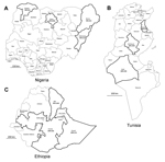
We found serologic evidence and supporting research for the circulation of Middle East respiratory syndrome coronavirus among dromedary camels in multiple African countries. Circulation of the virus among dromedaries across broad geographic areas of Africa may indicate that cases of this disease in humans are currently underdiagnosed outside the Arabian Peninsula.
A novel betacoronavirus (ΒCoV), Middle East respiratory syndrome coronavirus (MERS-CoV), was identified as the cause of severe respiratory disease in humans during 2012 (1). In August 2013, dromedary camels (Camelus dromedarius) were implicated for the first time as a possible source for human infection on the basis of the presence of MERS-CoV neutralizing antibodies in dromedaries from Oman and the Canary Islands of Spain (2). Since then, the presence of MERS-CoV antibodies in dromedaries has been reported in Jordan (3), Egypt (4,5), the United Arab Emirates (6,7), and Saudi Arabia (8,9). In October 2013, analysis of an outbreak associated with 1 barn in Qatar (10) found dromedaries and humans to be infected with nearly identical strains of MERS-CoV. Further proof of widespread circulation of MERS-CoV among dromedaries was provided by studies from Egypt and Saudi Arabia (5,9). These findings have raised questions about the geographic distribution of MERS-CoV among camel populations elsewhere. Here, we report our assessment of the geographic distribution of MERS-CoV circulation among dromedaries in Africa by serologic investigation of convenience samples from these animals in Nigeria, Tunisia, and Ethiopia.
full article
Dispatch
Geographic Distribution of MERS Coronavirus among Dromedary Camels, Africa
Chantal B.E.M. Reusken1Comments to Author , Lilia Messadi1, Ashenafi Feyisa1, Hussaini Ularamu1, Gert-Jan Godeke, Agom Danmarwa, Fufa Dawo, Mohamed Jemli, Simenew Melaku, David Shamaki, Yusuf Woma, Yiltawe Wungak, Endrias Zewdu Gebremedhin, Ilse Zutt, Berend-Jan Bosch, Bart L. Haagmans, and Marion P.G. Koopmans
Author affiliations: Netherlands Centre for Infectious Disease Control, Bilthoven, the Netherlands (C.B.E.M. Reusken, G.-J. Godeke, I. Zutt, M.P.G. Koopmans); Erasmus Medical Center, Rotterdam, the Netherlands (C.B.E.M. Reusken, B.L. Haagmans, M.P.G. Koopmans); National Veterinary Medicine School, University of La Manouba, Sidi Thabet, Tunisia (L. Messadi, M. Jemli); Addis Ababa University College of Veterinary Medicine and Agriculture, Bishoftu, Ethiopia (A. Feyisa, F. Dawo, S. Melaku, E. Z. Gebremedhin); National Veterinary Research Institute, Vom, Nigeria (H. Ularamu, A. Danmarwa, D. Shamaki, Y. Woma, Y. Wungak); Faculty of Veterinary Medicine, Utrecht University, Utrecht, the Netherlands (B.-J. Bosch); 1These authors contributed equally to this article.
Suggested citation for this article
Abstract
We found serologic evidence and supporting research for the circulation of Middle East respiratory syndrome coronavirus among dromedary camels in multiple African countries. Circulation of the virus among dromedaries across broad geographic areas of Africa may indicate that cases of this disease in humans are currently underdiagnosed outside the Arabian Peninsula.
A novel betacoronavirus (ΒCoV), Middle East respiratory syndrome coronavirus (MERS-CoV), was identified as the cause of severe respiratory disease in humans during 2012 (1). In August 2013, dromedary camels (Camelus dromedarius) were implicated for the first time as a possible source for human infection on the basis of the presence of MERS-CoV neutralizing antibodies in dromedaries from Oman and the Canary Islands of Spain (2). Since then, the presence of MERS-CoV antibodies in dromedaries has been reported in Jordan (3), Egypt (4,5), the United Arab Emirates (6,7), and Saudi Arabia (8,9). In October 2013, analysis of an outbreak associated with 1 barn in Qatar (10) found dromedaries and humans to be infected with nearly identical strains of MERS-CoV. Further proof of widespread circulation of MERS-CoV among dromedaries was provided by studies from Egypt and Saudi Arabia (5,9). These findings have raised questions about the geographic distribution of MERS-CoV among camel populations elsewhere. Here, we report our assessment of the geographic distribution of MERS-CoV circulation among dromedaries in Africa by serologic investigation of convenience samples from these animals in Nigeria, Tunisia, and Ethiopia.
full article
Comment